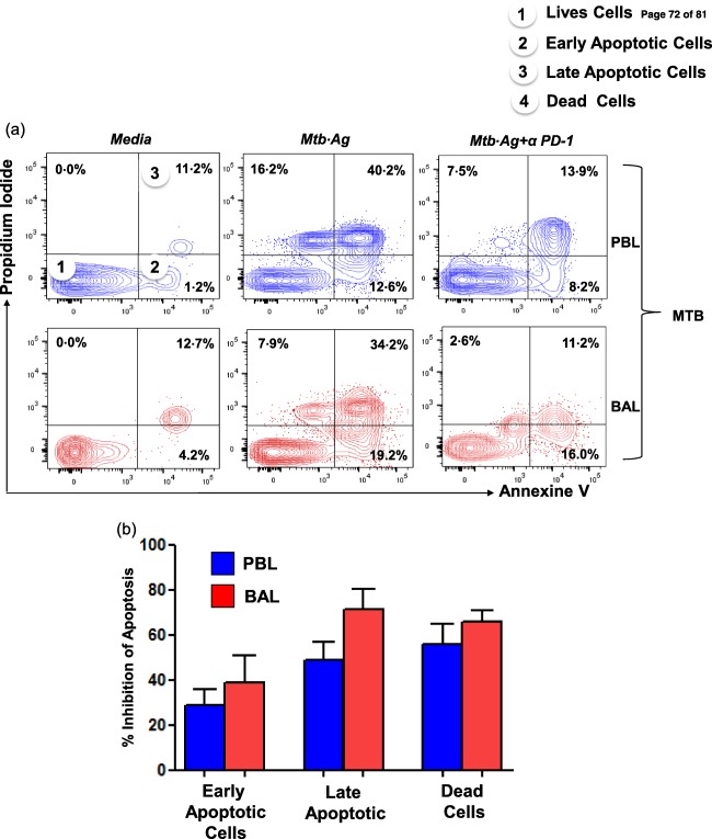
Figure 5.

Programmed death‐1 (PD‐1) blockade rescues CD3+ T cells from Mycobacterium tuberculosis‐induced apoptosis. Mononuclear cells obtained from peripheral blood (PB) and bronchoalveolar lavage (BAL) fluids from miliary tuberculosis (MTB) patients (n = 3) were cultured with M. tuberculosis antigen in the presence and absence of PD‐1 blockade for 24 h. Cultured cells were analysed for co‐expression of annexin V and propidium iodide (PI) on gated CD3+ T cells by fluorescence activated cell sorter (FACS). (a) Representative flow cytometry plots show the distribution of M. tuberculosis‐specific early apoptotic (annexin V+ only), late apoptotic (annexin V, PI dual‐positive) and dead cell (PI+ only) proportions under each experimental condition. (b) Inhibition of apoptosis following blocked PD‐1 was significantly higher in the BAL‐derived T cells. [Colour figure can be viewed at wileyonlinelibrary.com]
