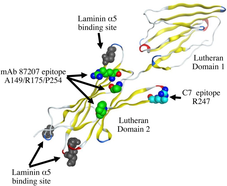
Fig 10. Mapping the amino acid residue recognized by C7 scFv on a three-dimensional model of Lu D1 and D2 domains.
An amino acid residue of the C7 scFv epitope mapped on the crystal structure of Lu D1 and D2 domains (Protein Data Bank code 2PET) [16]. The location of Arg247 is not very close to the epitope of the mAb87207 epitope. The binding sites of laminin α5 on Lu D2 domain are located on the opposite side of Arg247.

