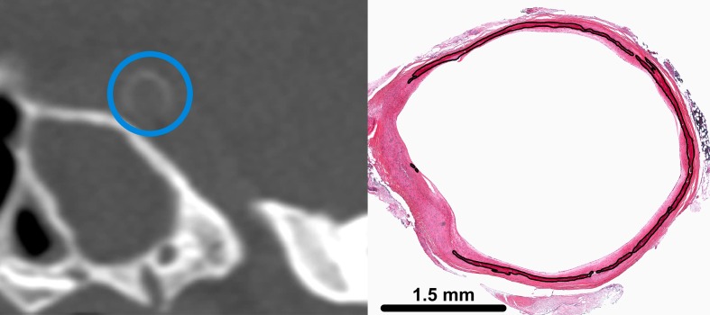
Fig 2. Internal elastic lamina calcification in the intracranial internal carotid artery (iICA) on a coronal brain CT image (left) and on a histological slide (right).
On CT a blue circle is placed around the iICA. Calcification area of the internal elastic lamina is indicated by the black line. Reprinted from A. Vos et al. Stroke. 2016;47:221–223 (Fig 1A) under a CC BY license, with permission of the American Heart Association, original copyright 2016 American Heart Association.

