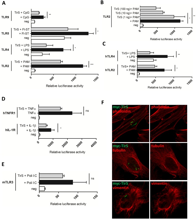
Fig 3. TirS interferes with TLR and IL-1R signaling.
(A) HEK293T cells were transiently transfected for 24 h with the luciferase reporter vector and murine TLR2, TLR4, TLR5, or TLR9 in the presence or absence of TirS (50 ng). Cells were then stimulated with the appropriate ligand (PAM, LPS, Fl-ST, and CpG) for 6 h before measurement of luciferase activity. White bars correspond to negative control, black bars to cells stimulated with the appropriate ligand, and gray bars to cells transfected with TirS and stimulated with the ligand. Data represent the means ± SEM of the relative luciferase activity and were obtained from duplicates of 3 independent experiments. (B) Luciferase activity of murine TLR2 following transfection with increasing amounts of vector encoding tirS (1, 50, 100 ng) in order to obtain different levels of expression and for (C) human TLR2 and TLR4. (D) Effect of TirS on TNFR and IL-1R activation following TNF-α or IL-1β stimulation, respectively and (E) TLR3 following stimulation with poli:IC. (F) HeLa cells were transfected with myc-TirS for 10 h and labeled for myc (green) and different components of the cytoskeleton: actin (top panels, phalloidin), microtubules (medium panels, tubulin), or intermediate filaments (bottom panels, vimentin). ns: not significant, * p < 0.05, ** p < 0.01, and *** p < 0.001.

