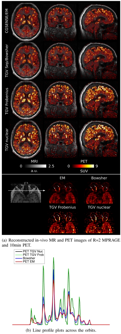
Fig. 8.
a) Overlays of MR and PET in a single transversal plane and coronal and sagittal reformats for the R=2 MPRAGE and 10min PET scan, comparing reference CG SENSE MR and a PET EM reconstruction (first row), Bowsher using separate TGV MR as the image prior (second row) and multi-channel joint MR-PET TGV reconstruction with Frobenius (third row) and nuclear (fourth row) norm coupling. A zoomed view of a region in the transversal plane depicting ocular muscles and orbital fat is shown in the third row. Sharpness and visibility of fine image features is clearly improved for the PET images with a multi-channel TGV regularizer. b) A line profile plot along the dashed arrow in the MR image of the orbital region. The orbital muscles show up as four distinct peaks (again highlighted by arrows), which show sharper separation from the surrounding fat region in the Joint TGV reconstructions.

