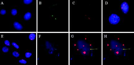
FIG. 1.
Detection of viral DNA and nascent transcription centers in CaSki and SiHa cervical cancer cell lines. (A and E) Detection of HPV-16 DNA (Cy3; red) in SiHa cells (A) or CaSki cells (E) with nick-translated probes in permeabilized interphase cells. (B and C) Simultaneous in situ detection in a single interphase SiHa cell of nascent HPV-16 RNA (FITC) (B) and HPV-16 DNA (Cy3) (C) by sequential hybridization with a nick-translated genomic probe. (D) Interphase chromosome analysis of SiHa cells for nascent HPV transcripts (FITC) and the chromosome 13 territory (Cy3). (F to H) Simultaneous in situ detection in a single interphase CaSki cell of nascent HPV-16 RNA (FITC) (F) and viral DNA (Cy3) (G) by sequential hybridization with a nick-translated genomic probe. (H) Merged image of panels F and G. Images were captured with a ×40 (A and E) or ×100 (B to D and F to H) objective lens but enlarged to different extent to display several cells (A and E) to show reproducibility or for a single cell for clarity (B to D and F to H). In all panels, nuclear DNA was stained with DAPI (blue).

