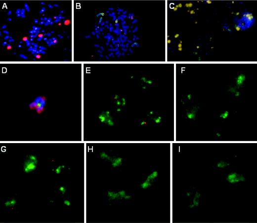
FIG. 2.
Mapping of the HPV transcription center in CaSki cells. (A) In situ hybridization to detect integrated HPV DNA (Cy3) in metaphase chromosomes (DAPI) in a CaSki cell. (B and C) Localization of HPV-16 DNA in CaSki cells by dual hybridization with a HPV-16 DNA probe (Cy3) and individual chromosome paints (FITC) on metaphase chromosome spreads (DAPI). A high-copy-number of HPV-16 DNA was found on two of the four major chromosomes 2 (green chromosomes with a yellowish arm in panel B). Chromosome 9 harbors no HPV DNA (C). (D) One or a few copies of HPV-16 DNA (FITC) were found on one of three acrocentric chromosomes 14 (Cy3). The paint did not color the entire metaphase chromosome (DAPI). (E to I) Interphase chromosome analysis of CaSki cells for nascent HPV transcription (Cy3) and chromosomal territory domains (FITC) showing chromosome 14 (E), chromosome X (F), chromosome 13 (G), chromosome 6 (H), and chromosome 22 (I). All images were captured with a ×100 objective lens but were enlarged to different extents for clarity.

