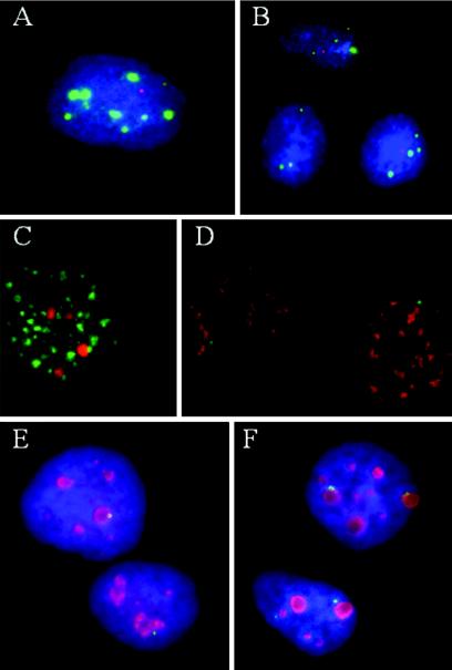
FIG. 8.
Perinucleolar localization of PVTD. With the exception of nucleoli, no association was found between PVTD, as denoted by nascent HPV RNA, and several nuclear domains, as specified. (A to E) CaSki cells; (F) SiHa cells. (A) ND10 domains detected with an antibody to PML in green and viral RNA in red; (B) p220NPAT, a Cajal body-associated protein in green and viral RNA in red; (C) SC35 domain in green and integrated HPV DNA in red; (D) SC35 domain in red and nascent HPV RNA in green; (E and F) antibody to C23 (nucleolin) in red and HPV RNA in green. All images were captured at ×100 magnification and are presented at a similar magnification except panel B, which is shown at a lower magnification.

