
Fig 1. Wnt signaling prevents Aβo-induced mitochondrial permeability transition pore opening in living neurons.
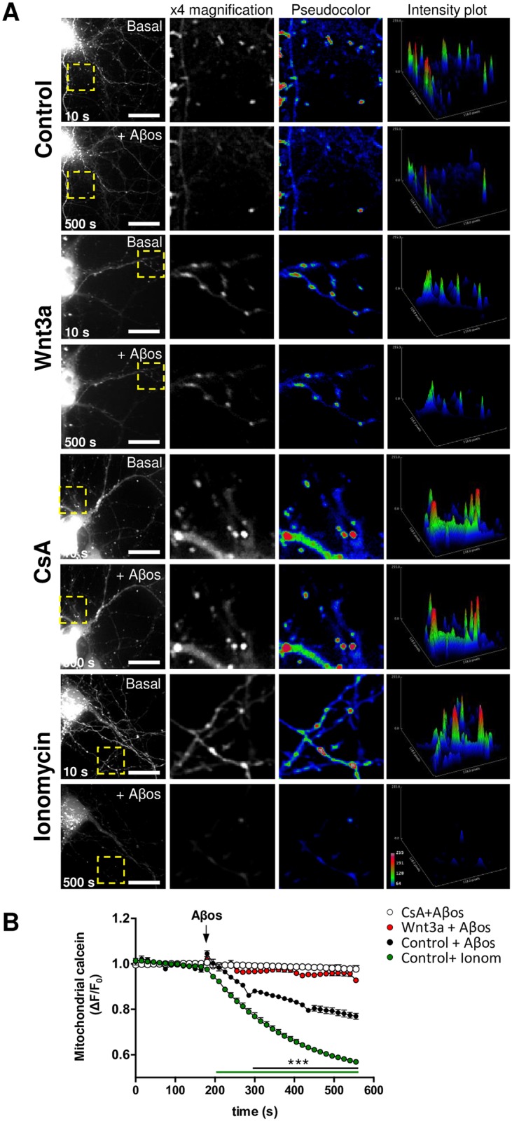
(A) Representative images showing DIV10 hippocampal neurons treated with control media or with recombinant Wnt3a protein (300 ng/ml) for 24 h and loaded with calcein-Co2+ to stain mitochondria. 20 μM CsA and 0.5 μM ionomycin were used as negative and positive controls for mPTP induction, respectively. The fluorescence intensity decay of mitochondrial calcein corresponds to mPTP opening in response to Aβos exposure. Yellow rectangles indicate magnified regions. Scale bar, 10 μm. Close-up photos are shown as 8-bit black and white images (x4 magnification) as well as pseudocoloured images. Intensity plots represent the fluorescence intensity profile of each magnified region. (B) Time-lapse quantification of the fluorescence intensity variations. The white horizontal bar on the graph indicates the addition of Aβos. The results represent the analysis of 5–7 neurites from 3–4 neurons per experiment. The graph shows the mean ± SEM of n = 7 independent experiments. Statistical analysis was performed with two-way ANOVA with post hoc Bonferroni correction: ***p<0.0005.