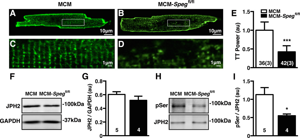
Figure 7. Loss of TTs and JPH2 phosphorylation precedes heart failure in MCM-Spegfl/fl mice.
(A–B) Representative images of ventricular myocytes isolated from MCM-Spegfl/fl mice or MCM controls 2 weeks post tamoxifen stained with di-8-ANEPPS and zoom (C–D) showing TT structure. (E) Quantification of TT Power. (F) Representative blot of total JPH2 with GAPDH as loading control from mouse heart lysates 2 weeks post tamoxifen and (G) corresponding quantification. (H) Representative image of JPH2 phosphoserine (pSer) Western blot normalized to JPH2 from immunoprecipitates from mice 2 weeks post tamoxifen and (I) quantification of phosphoserine normalized to total JPH2. Number in bars = number cells (mice); number hearts. * P<0.05.

