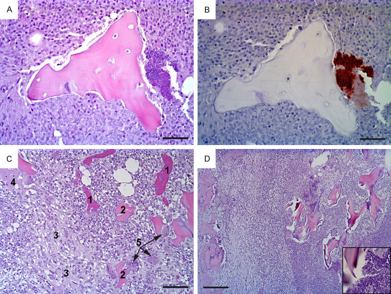
Figure 1.

Histopathology of osteomyelitis in pig A (pictures A-C) and pig E (picture D). Pig A: (A) shows the center of a bone lesion with necrotic trabecular bone surrounded by necrotic neutrophils (hematoxylin and eosin stain); at the right hand side of the figure is a colony of bacteria (blue) which in (B) are identified as S. aureus (brown) (immunohistochemistry); (C) shows the periphery of the lesion, disclosing blood vessels packed with erythrocytes (1), necrotic bone (2), fibroplasia (3), new bone formation (4) and osteoclasts (5) (hematoxylin and eosin). Pig E: (D) presents a similar lesion to that in in pig A, i.e. a subacute, suppurative, and necrotizing osteomyelitis, here bordering the cortex of the bone (left hand side) (hematocylin and eosin); insert is a close up of the bacteria seen in the necrotic center. Bar (A and B) = 25 μm, (C) = 50 μm, and (D) = 100 μm.
