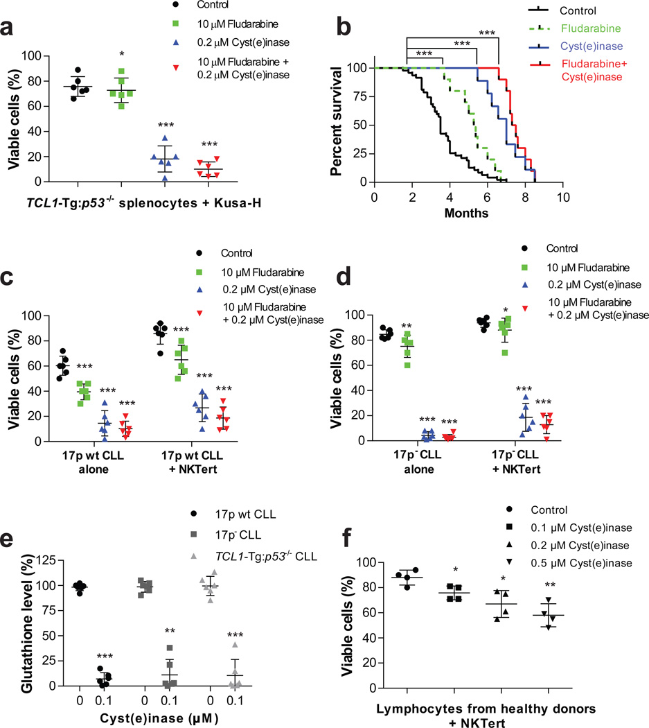
Figure 4. In vitro and in vivo efficacy of Cyst(e)inase in the TCL1-Tg:p53−/− mouse model and primary CLL cells.
(a) Cell viability 48 h after treatment with fludarabine, Cyst(e)inase, or their combination in splenocytes isolated from TCL1-Tg:p53−/− mice co-cultured with murine stromal Kusa-H cells (n = 6 per group). (b) Survival curve (Kaplan-Meier) of TCL1-Tg:p53−/− mice following either no treatment (n = 47); or treatment with fludarabine (n = 10); Cyst(e)inase (n=10); or their combination (n = 10). (c–d) Cell viability 48 h after treatment with fludarabine, Cyst(e)inase, or their combination in (c) primary 17p wt CLL cells; and (d) primary 17p- CLL cells either cultured alone or co-cultured with stromal NKTert cells (n = 6 different CLL patient samples per phenotype). (e) Relative GSH levels as assessed by spectrophotometric analysis 24 h after treatment with Cyst(e)inase in primary 17p wt, 17pCLL cells, and mouse splenocytes isolated from TCL1-Tg:p53−/− mice (n = 6 per group). (f) Cell viability 48 h after treatment with Cyst(e)inase in normal lymphocytes isolated from healthy donors co-cultured with NKTert cells (n = 4). For (a, c, d, f) cell viability was assessed by flow cytometry following double staining of cells with Annexin-V and PI. Data are expressed as mean ± s.d. (a, c–f). *P<0.05; **P<0.01; ***P<0.001; two-sided Student’s t-test (a, c–f) or log-rank (Mantel-Cox) test (b).

