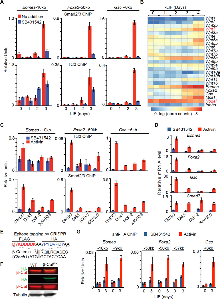
Figure 5. Wnt expression is rate limiting for the onset of mesendoderm differentiation.
A. ChIP-qPCR analysis of Smad2/3 and Tcf3 binding to the Eomes −10kb, Foxa2 −50kb and Gsc +6kb enhancers of EBs at the indicated times in LIF-free culture media. SB was added as indicated to inhibit signaling by endogenous nodal.
B. Heatmap of Wnt family mRNA expression levels (RNA-Seq) during d0 to d4 EB differentiation. Scale represents the log2 normalized read counts. Eomes, Foxa2, Gsc, Mixl1, Nodal and Inhba (encoding Activin A) expression levels are also presented for reference. Two biological replicates were analyzed for each condition.
C. ChIP-qPCR analysis of Tcf3 and Smad2/3 binding to the Eomes −10kb, Foxa2 −50kb and Gsc +6kb and enhancers in d3 EBs treated with SB or AC, and with DMSO or Wnt signaling inhibitors, Dkk1, IWP-2 or XAV939 as indicated.
D. Analysis of indicated mRNA levels (qRT-PCR) in d3 EBs that were incubated with SB or AC for 2 h. Wnt signaling inhibitors Dkk1, IWP-2 or XAV939, or DMSO vehicle were added as indicated.
E. Scheme of CRISPR mediated FLAG-HA epitope tagging for Ctnnb1/β-catenin N-terminus.
F. Western immunoblot analysis for β-cateninFLAG-HA (β-CatF-H) cell line that was inserted with FLAG-HA tags at Ctnnb1 N-terminus.
G. Anti-HA ChIP-qPCR analysis at Eomes −10kb and +9kb, Foxa2 −53kb, −50kb and −37kb, and Gsc +6kb enhancers.
See also Figure S5.

