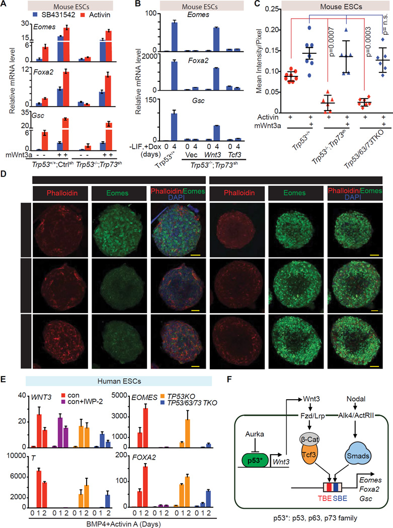
Figure 7. Wnt3 mediates p53 family action in mesendoderm specification.
A. qRT-PCR analysis of indicated mRNA levels in control ES cells (Trp53+/+;Ctrlsh), or p53/p73-depleted cells (Trp53−/−;Trp73sh), that were treated with or without recombinant mouse Wnt3a (mWnt3a) for 24h, followed by addition of SB or AC for 2 h.
B. qRT-PCR analysis of indicated mRNA levels in p53/p73-depleted cells with doxycycline (Dox)-inducible expression of Wnt3, Tcf3 or empty vector control. Assays were performed at d0 or day 4 of Dox treatment under differentiation conditions. Error bars represent s.e.m.
C. Quantitation of Eomes (green) signal in Panel C. Each dot represents the signal measurement from one EB. P value was calculated using Mann-Whitney test.
D. Immunofluorescence analysis of Eomes (green) in d3 EBs derived from the Trp53+/+;Ctrlsh, Trp53/63/73 TKO, or Trp53−/−;Trp73sh ES cells. Cells were treated with AC for 2h or AC plus mWnt3a for 20h. Scale bars, 50µm. Phalloidin (red) was used to stain F-actin and mark the cell boundaries.
E. qRT-PCR analysis of mRNA levels in control H1 hESC line with or without IWP-2 treatment, and in TP53KO and TP53/63/73 TKO derivative cell lines during BMP4 and AC induced differentiation.
F. Scheme of the p53 family-Wnt-Nodal network driving mesendoderm specification.
See also Figure S7.

