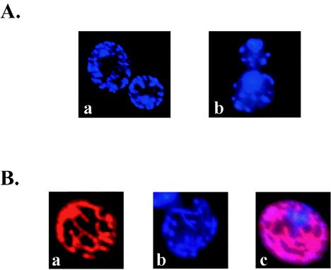
FIG. 2.
Distribution of DB75 within live yeast cells. Comparison of the fluorescent localization of DB75 and DAPI (A). UV fluorescent microscopy images of wet mounts of S. cerevisiae D273-10B cells treated for 1 h with either 7.1 μM DAPI (a) or 1.0 μM DB75 (b) were captured. The cellular distributions of DAPI and DB75 are similar, suggesting that DB75 accumulates within the nuclei and mitochondria of yeast cells. Fluorescent localization of MitoFluor Red, DB75, and MitoFluor Red plus DB75 (B). UV fluorescent microscopy images of wet mounts of S. cerevisiae D273-10B cells treated for 1 h with 25 nM MitoFluor Red 589 (a), 1.0 μM DB75 (b), or 25 nM MitoFluor Red 589 plus 1.0 μM DB75 (c) were captured. Fluorescent colocalization of MitoFluor Red with DB75, as indicated by pink fluorescence, confirms that DB75 enters yeast mitochondria.

