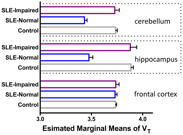
Figure 1.
Comparison of estimated marginal means of VT in cerebellum, hippocampus, and frontal cortex, obtained during the GLM multivariate analysis. Within patients with SLE, the cognitively impaired individuals (N = 4) showed higher radiotracer binding (mean regional VT) than cognitively normal patients with SLE (N = 6). Cognitively normal patients with SLE had diminished DPA binding compared to controls (N = 11). A pseudo-normalization was observed, going from cognitively normal to impaired in SLE, when compared to controls. Covariates appearing in the model are evaluated at the following values: VT_GM = 3.689, Age = 40.095.

