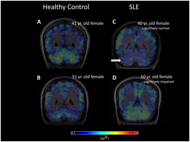
Figure 3.
Representative coronal view of DPA PET parametric (GMVT) images from age-matched control (A, B) and SLE (C, D) subjects. The cognitively normal SLE patient showed lower radiotracer binding in cerebellum (arrow in C) than the healthy control subject. Increased radiotracer binding in cerebellum was seen in the older, cognitively impaired SLE patient (B) when compared to C, whereas the opposite trend was suggested when comparing the younger (A) and older (B) healthy controls.

