
Fig. 2.
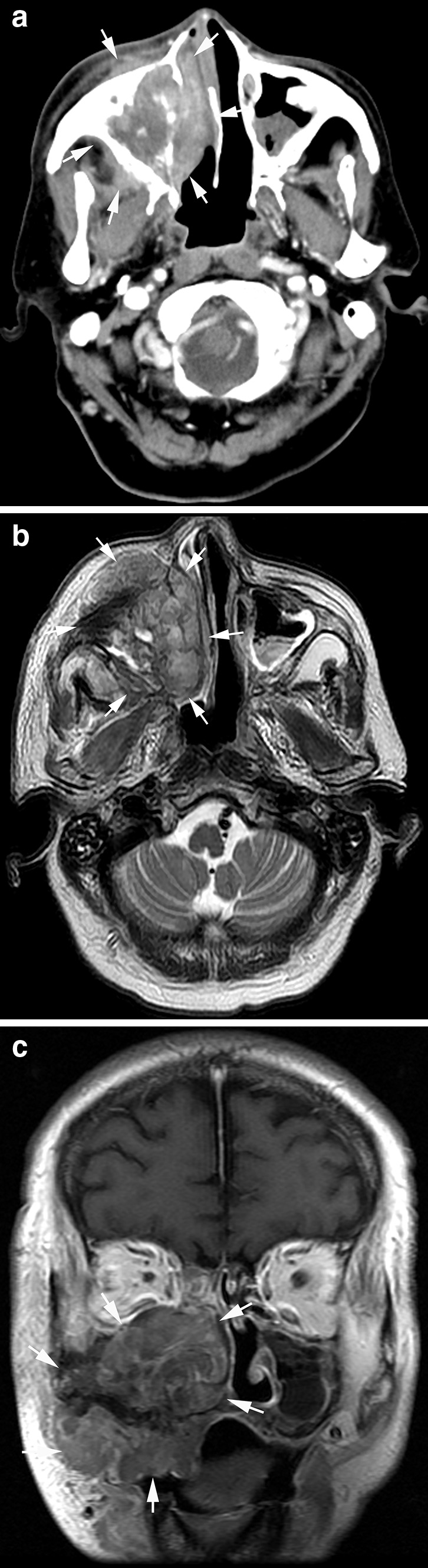
Radiation-induced osteosarcoma 17 years after radiotherapy of a 58-year-old man. a Axial contrast-enhanced CT image showed a large heterogeneous tumor in the right maxillary sinus invading the nasal cavity (arrows); b axial T2-weighted image showed heterogeneously high T2 signal intensity of the tumor; c coronal T1-weighted contrast-enhanced image showed the mildly enhanced tumor with wide invasion into the right maxillary region (arrows)