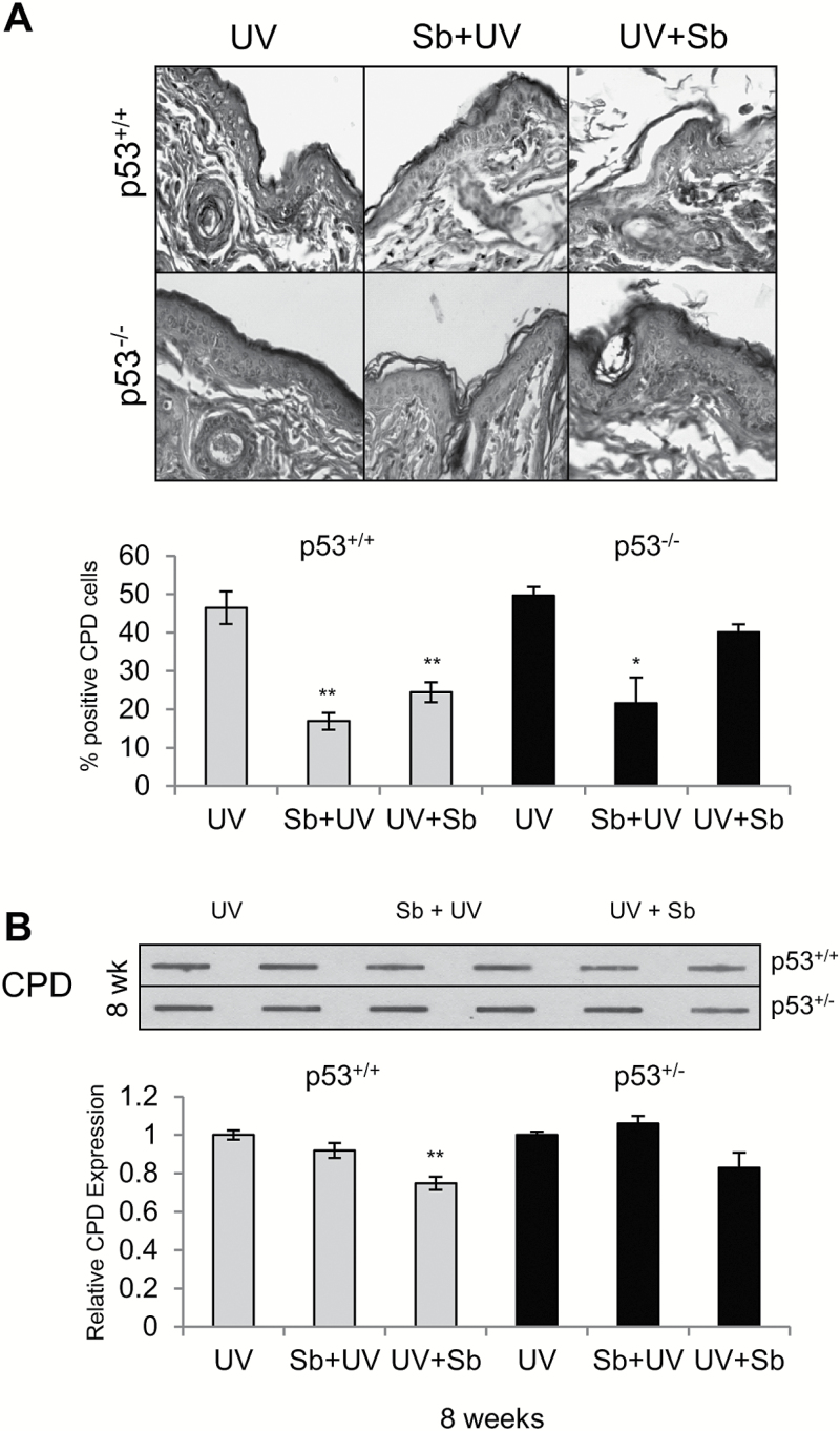
Figure 4.
Effect of silibinin treatment on DNA damage removal in SKH-1 p53+/+, p53+/− and p53−/− mice following UVB exposure. (A) p53+/+ and p53−/− mice were exposed to UVB with or without topical application of silibinin (9 mg in 200 µl acetone) prior to UVB (Sb + UV) or immediately after UVB exposure (UV + Sb) as detailed in Materials and Methods. 12 h following UVB exposure, skin was analyzed for CPDs by IHC as detailed in methods. The data shown in the bar diagram represents mean ± SEM of four samples. Representative pictures of CPD staining are presented below the bar diagram. (B) p53+/+ and p53+/− mice were exposed to UVB alone (5 days/week) or treated with 9 mg silibinin pre- or post-UVB treatment. After 8 weeks, skin was analyzed for CPD levels by Slot Blot method. The bar diagram represents the densitometric analysis of the CPD levels which is mean ± SEM of two independent experiments. **P < 0.001.

