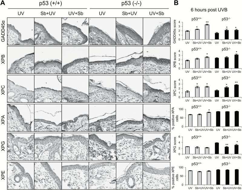
Figure 5.
The effect of silibinin treatment on UVB-induced GADD45α and NER pathway molecules in p53+/+ and p53−/− mouse skin following single UVB exposure. Mice were exposed to UVB (180 mJ/cm2) with or without topical application of silibinin (9 mg in 200 µl acetone) prior to UVB (Sb + UV) or immediately after UVB exposure (UV + Sb) as detailed in Materials and Methods. Six hours later, mice were killed and skin was collected and analyzed by IHC. (A) IHC staining for GADD45α, XPB, XPC, XPA, XPG and XPE. Representative photomicrographs are show at 400× magnification. (B) Relative percentage of stained cells or score for each IHC staining. The data shown in the bar diagram represents mean ± SEM of five samples. *P < 0.05 and **P < 0.001 versus respective UV irradiated group.

