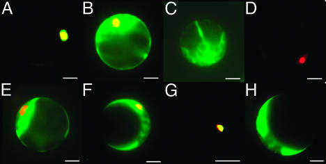
Fig. 4.
Subcellular localization of CPL1 and CPL2. TAP-GFP-CPL1 (A), TAP-GFP-CPL2 (B), TAP-GFP (C), GFP-CPL11-150 (E), GFP-CPL1151-639 (F), GFP-CPL1640-967 (G), and GFP (H) were transiently expressed in Arabidopsis protoplasts. NLSSV40-DsRed was used as a positive control for nuclear localization (D) and was coexpressed with GFP-fusion constructs (A-C and E-G). Fluorescent signals from GFP and DsRed were obtained by using standard FITC and rhodamine filter sets. The pictures show pseudocolor overlap images of GFP (green) and DsRed (red). Yellow indicates colocalization of GFP and RFP fusion proteins. (Scale bars: 10 μm.)

