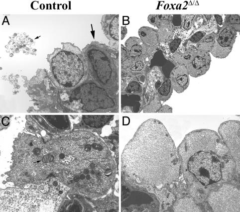
Fig. 2.
Lung ultrastructure in Foxa2Δ/Δ mice. Electron microscopy was performed on lungs from E18.5 Foxa2Δ/Δ (B and D) and littermates (A and C). Squamous type I cells (thick arrow) and cuboidal type II cells (thin arrow in C) containing lamellar bodies, apical microvilli, and highly organized rosette glycogen were observed in lungs of control mice (A and C). The densely stained cell below the type I cell is a capillary endothelial cell creating the gas-exchange area (A). Type I cells were not observed in the lungs of Foxa2Δ/Δ mice. The pulmonary vascular bed in Foxa2Δ/Δ mice was intact, but in the absence of type I cells, the thin-walled alveolar-capillary interface seen in the control mice was not formed. Type II cells were immature, lamellar bodies were absent in Foxa2Δ/Δ mice, apical microvilli were sparse, and particulate glycogen was absent in Foxa2Δ/Δ mice. Thin arrow (A) shows secreted surfactant. The micro-graphs are representative of two Foxa2Δ/Δ and littermate controls.

