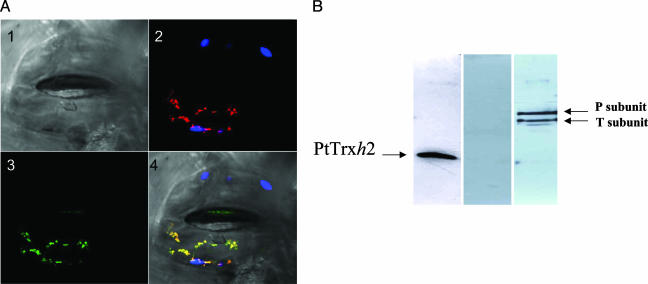
Fig. 1.
Mitochondrial localization of PtTrxh2. (A) Mitochondrial localization of the PtTrxh2 fused to enhanced GFP in N. tabacum stomata. (Image 1) Image of stomata from Nicotiana benthamiana under visible light. Only the lower guard cell was transfected. (Image 2) Autofluorescence of chlorophyll (blue) and fluorescence of the mitochondrial marker (red). (Image 3) Fluorescence of the fusion protein (green). (Image 4) Merged images. (B) Immunodetection of PtTrxh2 (Left), methionine sulfoxide reductase A (Center), and glycine decarboxylase complex subunits P and T (Right) in poplar purified mitochondria.

