
Fig. 1.
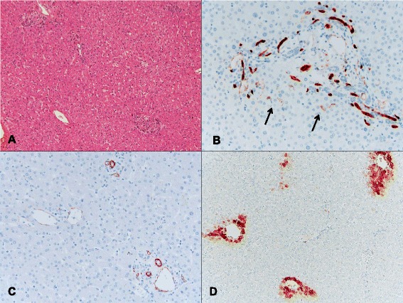
An example of a control group (case 29 in Table 5) in which a patient with CLRM did not receive chemotherapy prior to resection. a This case showed no significant sinusoidal dilatation, congestion, or parenchymal nodularity (H&E, ×40). b An immunohistochemical stain for CD34 showed the normal staining pattern with only focal weak sinusoidal staining around the portal tracts (arrows, ×200). c Positive immunoreactivity for smooth muscle actin (SMA) was seen in the portal blood vessels, bile ducts, and central venules. No SMA-positive stellate cells were present in the lobules (×200). d An immunohistochemical stain for GS revealed a normal perivenular staining pattern (×100)