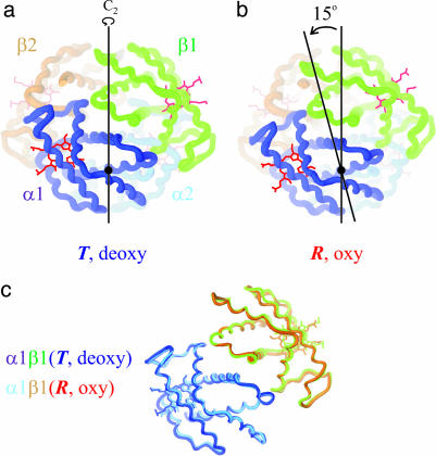
Fig. 1.
Schematic structures of hemoglobin made with the program ribbons. Shown are deoxyhemoglobin in the T quaternary structure (a) (Protein Data Bank ID code 4HHB) and oxyhemoglobin in the R quaternary structure (b) (Protein Data Bank ID code 1HHO). The quaternary conformational change consists of a 15° relative rotation of the αβ dimers that are interchanged by the twofold symmetry (C2) axis. (c) A comparison of the tertiary structures, using the invariant interface between α and β subunits of the αβ dimer as a reference (37), is shown.

