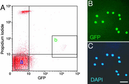
Fig. 1.
Purification of CR cells by FACS. (A) GFP-positive, PI-negative cells (fraction b) and GFP-negative, PI-negative cells (fraction a) at P2 were sorted by FACS as CR cells and non-CR cells, respectively. The PI fluorescence of viable CR cells was slightly higher than that of viable non-CR cells because of a slight overlap of GFP fluorescence in PI fluorescence measurement. (B and C) Purified GFP-positive CR cells were stained with 4′,6-diamidino-2-phenylindole (DAPI) and detected with microscopy. (Scale bar, 50 μm.)

