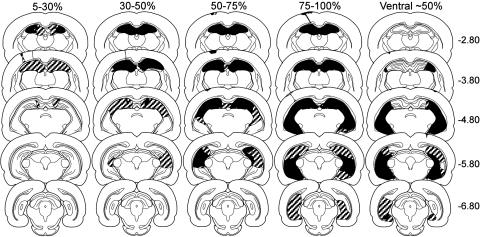
Fig. 1.
Reconstructions of coronal sections through the hippocampus showing the smallest (black) and largest (stippled) lesion for each of the four hippocampal lesion groups (damage extending from the dorsal hippocampus to include 5-30%, 30-50%, 50-75%, and 75-100% of total hippocampal volume) from experiment 1 and the ventral lesion group (damage to ≈50% of total hippocampal volume) from experiment 2. Note that the locus and extent of hippocampal damage for the 50-75% and 75-100% groups were similar in experiments 1 and 2 (experiment 2 not shown). All rats sustained bilateral damage to the CA cell fields and dentate gyrus. In cases where the lesion was not complete at a particular level of the dorsal hippocampus, the sparing was typically restricted to the most medial aspect of the dentate gyrus or CA1 cell field. There was no evidence of damage to the amygdala or perirhinal cortex. Numbers (right) represent the distance (mm) posterior to bregma. For an additional description of the lesions, see Supporting Text.

