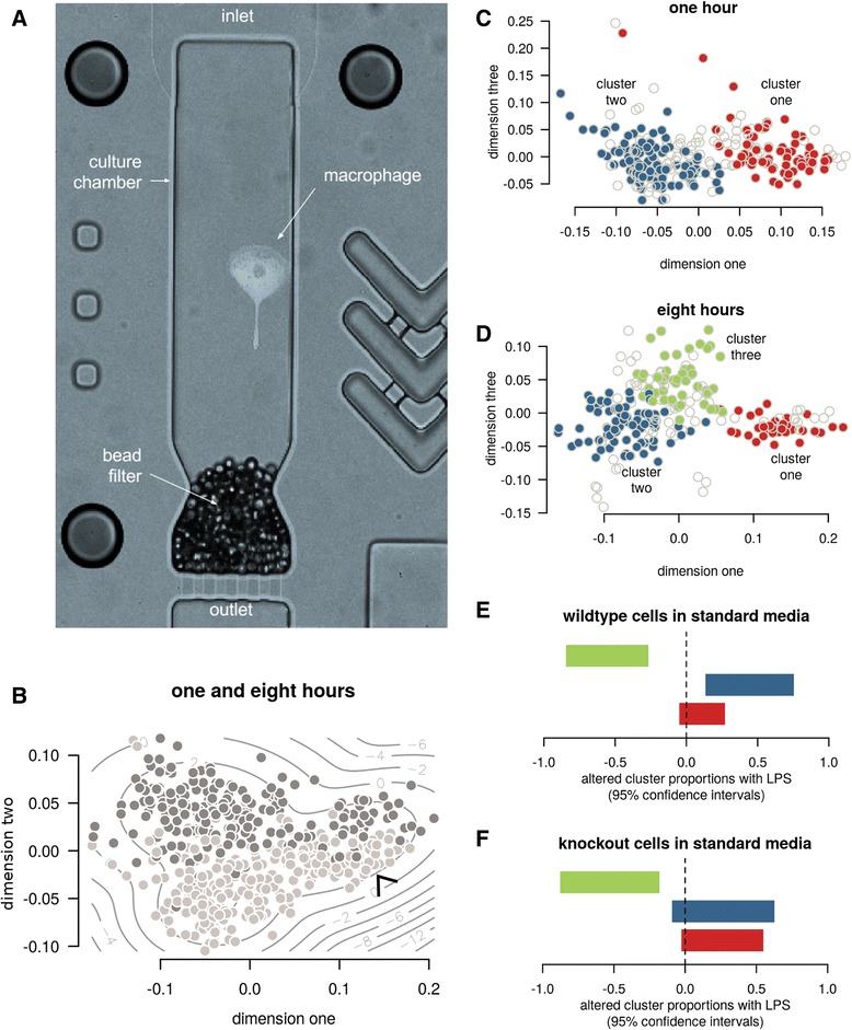
Fig. 1.

Macrophage culture and subtypes. a A single micro-volume culture chamber from the Polaris™ microfluidics chip, containing a macrophage. The media conditions per chamber can be modified to study microenvironmental perturbations. b Visualization of the major cell differences — such as with this multi-dimensional scaling of the transcriptomic rank correlations — demonstrated the separation of cells cultured for one hour (light), eight hours (dark), and a reproducible subcluster present across both time points (arrow). c&d Formal clustering confirmed this subcluster (cluster one) in addition to a third subcluster emerging after eight hours. The inner 50% of cells in each cluster are shown in colour for dimensions one and three to better convey relative cluster positions and densities. e&f Cluster three significantly reduced its proportion in the context of LPS and standard media. 95% confidence intervals for change in proportions are shown
