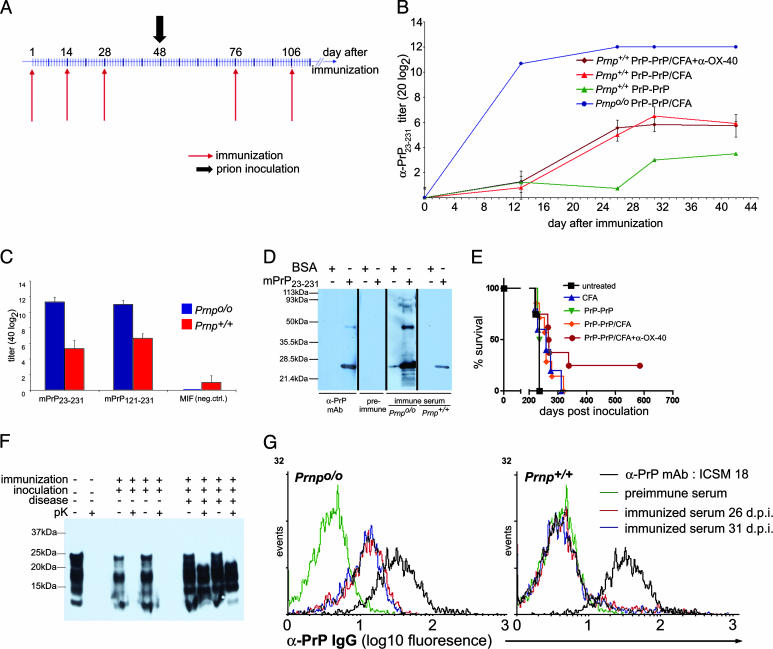
Fig. 1.
Immunization of Prnp+/+ mice induces anti-PrPREC but not anti-PrPC responses and is not protective. (A) Mice were immunized three times with PrP-PrP in 2-week intervals. Mice were then inoculated with prions i.p. (black arrow) and boosted with PrP-PrP every month until the first clinical signs of scrapie were recorded. (B) Antibody titers to PrPREC were investigated by ELISA. Upon PrP-PrP immunization, Prnpo/o mice developed early and high anti-PrPREC titers, whereas Prnp+/+ mice showed much lower titers at days 26–32 after immunization. (C) Specificity of measured titers. ELISA plates were coated with recombinant mouse PrP (mPrP23–231), a truncated variant of it (mPrP121–231), or an unrelated recombinant protein produced by a similar method in E. coli. The results indicate that antibodies were indeed directed to PrP rather than residual bacterial contaminants. MIF, Macrophage inhibitory factor. (D) Western blot using preimmune and immune serum of Prnpo/o and Prnp+/+ mice and monoclonal anti–PrP antibody 6H4 for control. All strips were incubated with the same α-mouse IgG secondary antibody to allow comparison. Immune serum of both immunized mice specifically recognized PrPREC, but not BSA, which was loaded in the same amount as a negative control. Preimmune serum of the same Prnp+/+ mouse showed no reactivity to PrPREC.(E) Survival plot visualizing incubation times until development of terminal disease of immunized Prnp+/+ mice after i.p. challenge with prions. Two immunized mice that received anti-OX-40 did not develop scrapie symptoms until 585 days postinoculation, when they were killed. (F) Western blot analysis of brain homogenates of prion-inoculated immunized mice. The two mice that displayed no signs of disease after >580 days postinoculation showed no PrPSc accumulation in the brain. (G) Flow cytometric analysis of sera from immunized mice on tg33 transgenic splenocytes with PrPC overexpression restricted to T cells. Cells were gated for live splenocytes and subsequently for CD3+ T cells. Histograms represent the intensity of the binding of anti-PrP antibodies present in sera of immunized mice. Serum of Prnpo/o mice at days 26 or 31 after immunization showed specific recognition of native PrPC (Left). Instead, sera of immunized WT mice and preimmune sera did not recognize native PrPC (Right).

