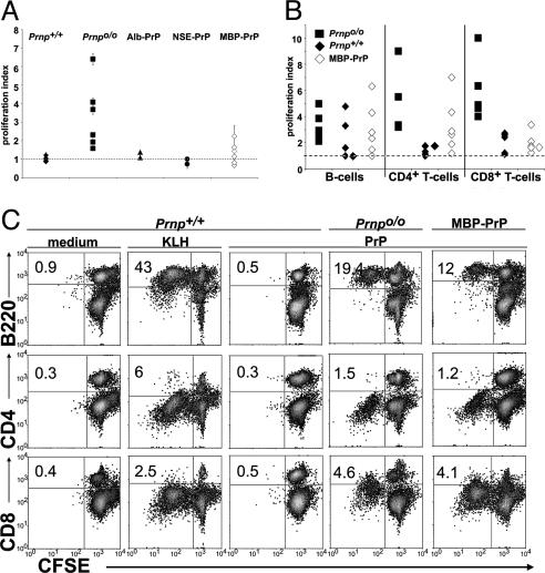
Fig. 4.
Cellular responses to PrPREC correlate with humoral responses. (A) Thymidine incorporation into lymph node cells 48 h after in vitro restimulation with PrPREC. Lymph node cells were taken from mice that had been immunized with PrPREC in CFA 7 days earlier and were cultured for 48 h in the presence or absence of PrPREC. The proliferation index represents the ratio of thymidine incorporation of antigen-stimulated to unstimulated cells. Each symbol summarizes triplicate cultures from one individual mouse (results from three independent experiments). The dotted line represents a ratio of 1 and corresponds to the thymidine incorporation of unstimulated cells. (B) CFSE stains from lymph node cells of immunized Prnpo/o, Prnp+/+, and MBP-PrP mice 4 days after in vitro restimulation with PrPREC. Prnp+/+-immunized mice showed poor or no proliferation of B220+ (B cells) or CD8+ (cytotoxic T cells) and totally unresponsive CD4+ (T helper cells), whereas Prnpo/o and MBP-PrP mice showed comparable proliferation in B and CD4+ T cells. CD8+ T cells were only moderately proliferative on some of the MBP-PrP mice. Proliferation index represents the ratio of the percentage of proliferating PrPREC-stimulated cells (lower CFSE intensity) to that of unstimulated cells. Results are of two independent experiments (Prnpo/o and Prnp+/+ n = 5, MBP-PrP n = 6). (C) CFSE stains from lymph node cells of immunized Prnpo/o, Prnp+/+, and MBP-PrP mice 4 days after in vitro restimulation with the antigen (PrPREC or KLH). One representative mouse of each genotype is depicted.

