Fig. 2.
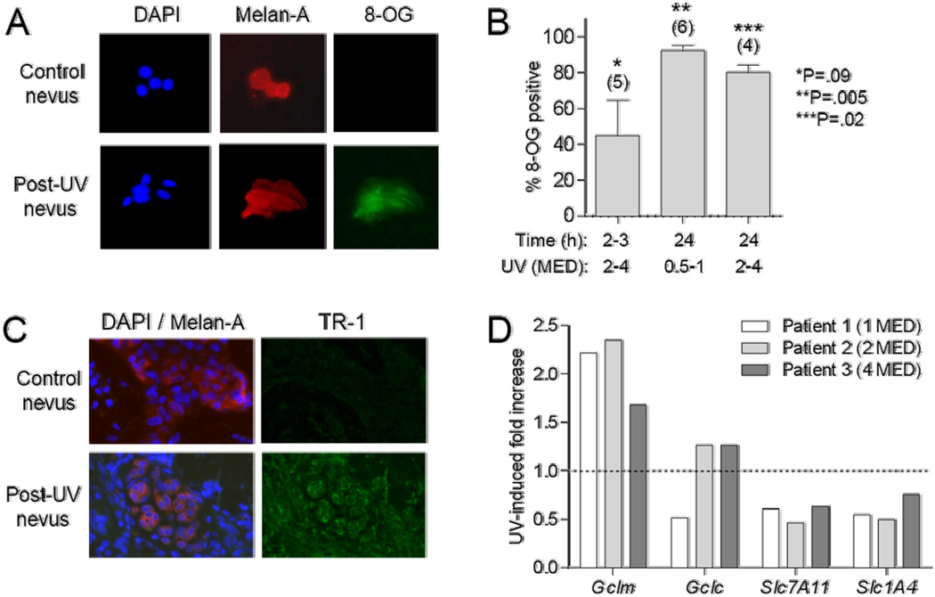
UV-induced markers of oxidative stress. A, Representative staining of dissociated nevus cells for DAPI, Melan-A, or 8-OG. B, Shown are percent nevus melanocytes positive for 8-OG, at indicated time and UV dose ranges. Error bars indicate SEM. Numbers of patients in each group indicated in parentheses. P values for comparisons of values for irradiated vs. unirradiated tissues at each condition determined by paired t tests. C, Representative staining of nevus sections for DAPI, Melan-A, and TR-1. D, Expression of Gclm, Gclc, Slc7A11, and Slc1A4 genes from RNA isolated from control and UV-irradiated nevi 24h after SSR exposure (1–4 MED) in three subjects. Data expressed as ratio of normalized signals from UV-treated to control nevi.

