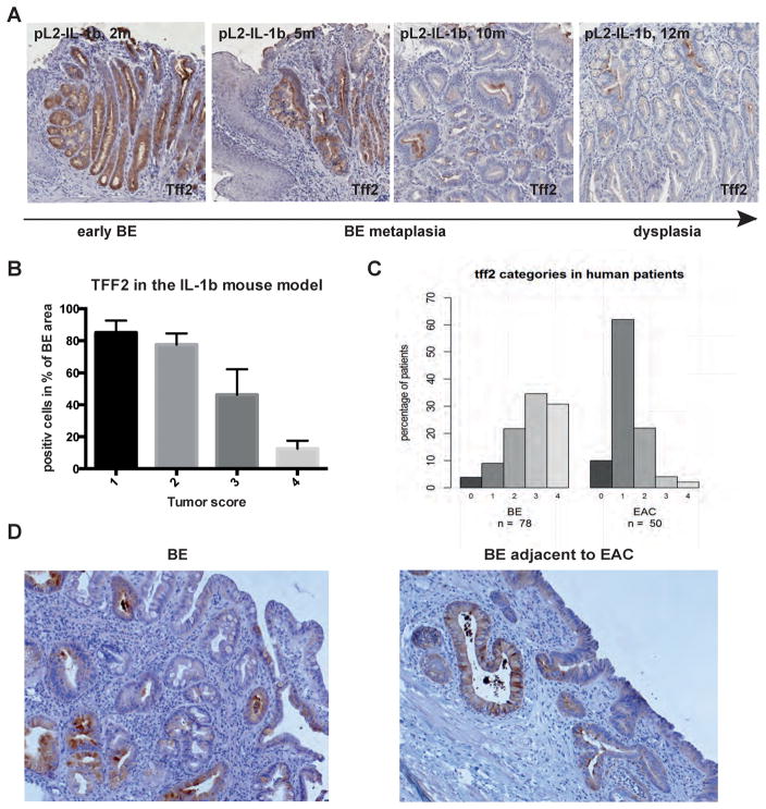
Figure 2.
A) TFF2 immunohistochemistry of BE metaplasia in the pL2-IL-1β mouse model with increasing status of early (2 months) to metaplastic (5 and 10 months) BE and dysplasia (12 months). B) Histopathologcial score in correlation with TFF2 staining in the pL2-IL-1β mouse model (ANOVA p<0.0001, R square 0.8728, F 54.91). C) TFF2 staining in human biopsies from BE or adjacent to EAC tissue was scored from 0 to 4, percentage of each score in both groups is shown (4/9/22/35/31% in BE as well as 10/62/22/4/2% in EAC). D) Representative TFF2 staining of human biopsies from BE or adjacent to EAC tissue.

