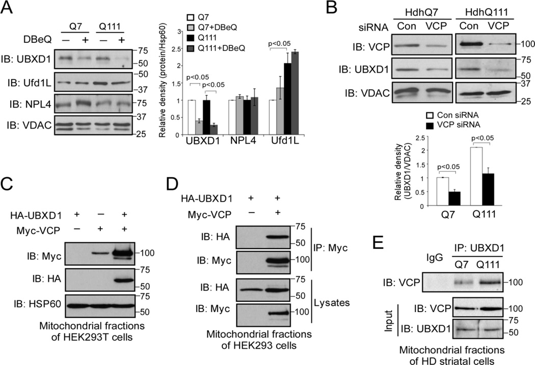
Figure 4. UBXD1 is a VCP mitochondrial cofactor.
(A) HdhQ7 and HdhQ111 cells were treated with VCP inhibitor DBeQ (10 µM) for 16 hours. Western blot analysis of mitochondrial fractions was performed with the indicated antibodies. The data are mean ± SE of three independent experiments. *, p<0.05 vs. Q111 cells. ANOVA with post-hoc Holm-Sidak test. (B) HdhQ111 cells were transfected with control or VCP siRNA for 48 h. Mitochondrial VCP and UBXD1 protein levels were detected with anti-VCP and anti-UBXD1 antibodies by WB. The data are mean ± SE of three independent experiments. *, p<0.05 vs. cells with con siRNA. ANOVA with post-hoc Holm-Sidak test. (C) Control vector or Myc-VCP was co-expressed with HA-UBXD1 in HEK293T cells for 36 hours. Western blot analyses of mitochondrial fractions were performed with anti-Myc, anti-HA, and anti-HSP60 antibodies. (D) HEK293T cells were co-transfected with Myc-VCP and HA-UBXD1 for 36 hours. Immunoprecipitates (IP) with anti-Myc were immunoblotted (IB) with anti-HA antibody. (E) Mitochondrial fractions were isolated from HdhQ7 and HdhQ111 cells. IP with anti-UBXD1 followed by IB with anti-VCP antibody was conducted. All blots shown are from three independent experiments.

