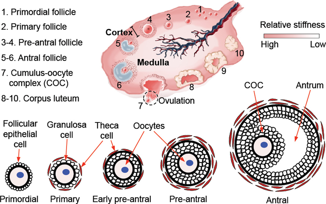
Figure 1.
A schematic illustration of the mammalian ovary showing the difference in mechanical properties between the ovarian cortex and medulla, the morphology of ovarian follicles at various developmental stages, and the ovulation of cumulus-oocyte complex (COC) from antral follicles leaving behind the corpus luteum. The schematic of mammalian ovary is reprinted and redrawn from reference [47] with permission from Elsevier.

