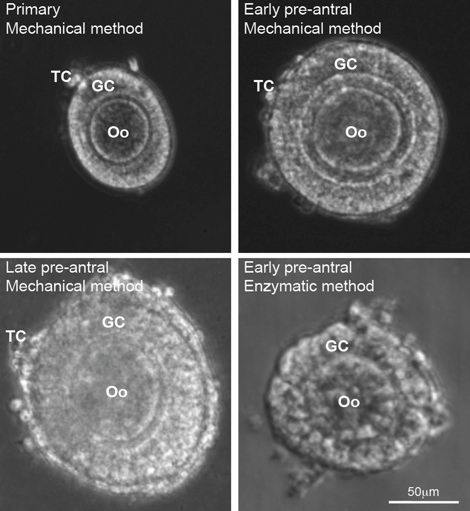
Figure 2.
Images showing the typical morphology of primary (75–99 µm), early preantral (100–125 µm), and late preantral (126–180 µm) follicles retrieved from ovaries of Peromyscus using the mechanical method together with that of early preantral follicle obtained using the enzymatic method: The follicles retrieved by the mechanical method have an intact outer membrane of theca cells (TCs) and an intact layer(s) of granulosa cells (GCs) in the middle together with a primary oocyte (Oo) in the center. In contrast, the middle and particularly, the outer layer of the follicles retrieved by the enzymatic method were severely compromised. The figure is reprinted and redrawn from reference [46] with permission from Mary Ann Liebert Inc.

