Figure 8.
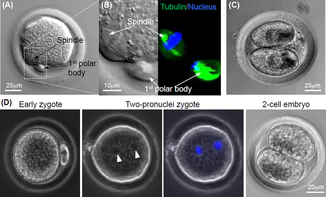
Quality of MII oocytes obtained by In vitro maturation of cumulus-oocyte complex from antral follicles developed from early preantral follicles by in vitro culture: (A) Typical image of a MII oocyte obtained from the antral follicles showing the characteristic 1st polar body and mitotic spindle, (B) nuclear and tubulin stains for visualizing the 1st polar body and mitotic spindle in the MII oocyte, (C) image of a two-cell embryo developed from the MII oocyte after parthenogenetic activation, and (D) typical micrographs showing development of MII oocyte after in vitro fertilization (early zygote) to the two-pronuclei and two-cell stages. The two pronuclei visible in the phase contrast image (arrow head) were further confirmed using fluorescence stain of the pronuclei (blue stains). The figure is reprinted and redrawn from reference [48] with permission from John Wiley & Sons, Inc. (for panels A–C) and from reference [46] with permission from Mary Ann Liebert Inc. (for panel D).

