Fig. 1.
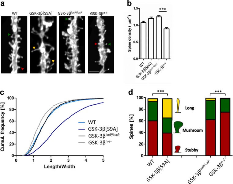
GSK-3β imbalance in neurons alters dendritic spine density and morphology. a Example photographs of DiI-stained secondary apical dendrites of granule neurons in the dentate gyrus in GSK-3β[S9A] and GSK-3βn−/− mice. Scale bar = 2 μm. b Spine densities of dentate gyrus neurons in GSK-3β[S9A] and GSK-3βn−/− mice. The data are expressed as mean ± SEM. ***p < 0.001 (Mann-Whitney test). c Cumulative histogram of dendritic spine length-to-width ratio in GSK-3βn−/− and GSK-3β[S9A] mice (p < 0.001, vs. GSK-3βloxP/loxP and WT; nested analysis of variance). d Spine morphology in GSK-3β[S9A] and GSK-3βn−/− mice. ***p < 0.001 (χ 2 test). GSK-3β[S9A]: n = 6; WT: n = 6; GSK-3βn−/−: n = 4; GSK-3βloxP/loxP: n = 5 mice)

