. 2016 Oct 8;13(6):443–452. doi: 10.21873/cgp.20007

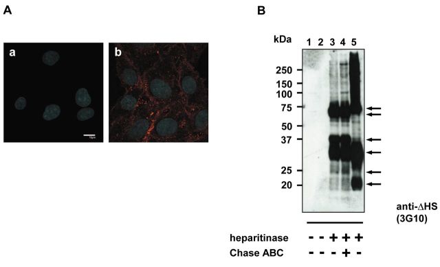
Figure 1. High levels of HSPGs expression can be detected in C6 glioma cells. The C6 glioma cell line was analyzed for expression of HSPGs. (A) Immunocytochemistry was performed using heparan sulfate-specific antibodies. C6 cells were cultured, fixed with PFA, and stained with 10E4 antibody (b) or with secondary antibodies only (a). The nuclei were stained with Hoechst 33342. Scale bars: 10 μm. (B) Proteoglycans isolated from confluent C6 glioma cells were purified on Q-Sepharose, incubated with or without heparitinase and immunoblotted with anti-ΔHS antibodies (3G10). Lanes: 1, digestion buffer only; 2, 3, 4 correspond to purified material from C6 glioma cells; 5, positive control for immunoblotting representing rat parathyroid proteoglycans.

Figure 1