. 2016 Oct 8;13(6):443–452. doi: 10.21873/cgp.20007

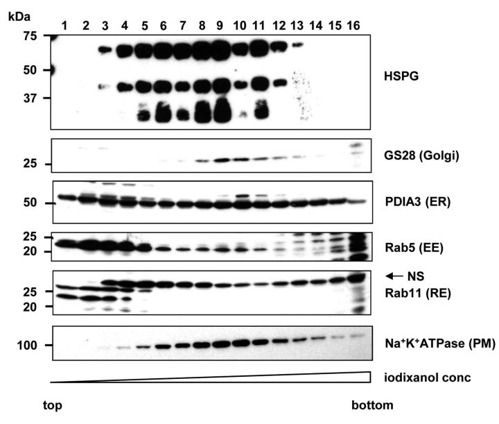
Figure 3. Subcellular fractionation of C6 glioma cells using OptiPrep ultracentrifugation gradient. C6 glioma cells were subjected to subcellular fractionation as described in the Materials and Methods section. Each fraction was analyzed by immunoblotting with specific antibodies for organelle markers and heparan sulfate, respectively. For organelle marker detection, equal volume of each fraction (14 μl/lane) was applied on gel. Depending on the analyzed marker, the samples were run under non-reducing (Na+-K+-ATPase, PIDA3 and Rab11) or reducing (GS28, Rab5 and HSPGs) conditions. To detect HSPGs, 90 μl of each sample was concentrated and treated with heparitinase prior the electrophoresis. Lanes: 1-16 correspond to subcellular fractions collected from the top of the gradient. In Rab11-stained membrane, bands marked with (NS) correspond to non-specific staining also described by manufacturer in antibody data sheet.

Figure 3