Figure 1.

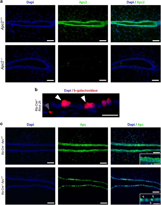
Deletion of the Apc proteins from murine mammary epithelium. (a) Mammary gland sections from Apc2+/+ and Apc2−/− mice were labeled for Apc2 using fluorescent IHC. Although Apc2 is expressed in Wt epithelium, Apc2−/− glands displayed comprehensive Apc2 loss (scale bar, 50 μm). (b) Virgin mammary gland sections from 10-week-old Blg-Cre+Rosa26+ mice were labeled for β-galactosidase to assess Cre-mediated recombination. Recombination occurred in a heterogeneous manner, primarily within luminal cells (white arrowheads) but occasionally detectable in apparent non-luminal cells (gray arrowhead) (scale bar, 25 μm). (c) Labeling of Virgin mammary glands section from 10-week-old Blg-Cre+Apcfl/fl mice for Apc using fluorescent IHC revealed a heterogeneous pattern of loss in epithelial cells (scale bar, 50 μm in main image, 25 μm in inlay).
