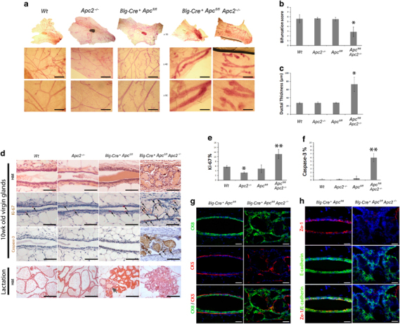
Figure 2.

Either Apc or Apc2 is dispensable, however, concurrent loss results in a range of epithelial disruptions. (a) Carmine alum-stained whole mount glands from 10-week-old virgin mice reveals that loss of either Apc protein alone is tolerated, whereas combined loss results in severe defects in ductal branching and epithelial thickening (scale bar, 200 um). (b) Quantification of ductal branching. (c) Quantification of epithelial thickening. (d) H&E-stained sections of mammary tissue from each genotype reveal epithelial disruptions with intraluminal ghost cell nodules in glands deficient for both Apc proteins (Blg-Cre+Apcfl/flApc2−/−) (scale bar, 50 μm). Labeling for both Ki-67 and caspase-3 exposed an increase in positive cells in epithelium deficient for both Apc proteins (arrows indicate positively labeled cells, scale bar, 50 μm). H&E-stained sections of lactating glands from each genotype revealed Apc2-deficient glands to be indistinguishable from Wt, Blg-Cre+Apcfl/fl glands displayed attenuated alveolar formation with occasional ghost cell nodules (arrow) and Blg-Cre+Apcfl/flApc2−/−glands displayed a complete lack of differentiated alveoli and vastly perturbed tissue architecture (scale bar, 50 μm). (e) Quantification of Ki-67-positive cells revealed a reduction in proliferation in Apc2−/− compared with Wt epithelium. A statistical increase was noticed in epithelium deficient for both Apc proteins versus all other genotypes (error bars, s.d., *P⩽0.05 versus Wt, **P⩽0.01 versus all other genotypes, Mann–Whitney U-test, n⩾3). (f) Quantification of caspase-3-positive cells revealed a statistical increase in apoptosis in epithelium deficient for both Apc proteins (error bars=s.d., **P⩽0.001 versus all other genotypes, Mann–Whitney U-test, n⩾3). (g) Sections of 10-week-old virgin glands from Blg-Cre+Apcfl/fl and Blg-Cre+Apcfl/flApc2−/−were double labeled for cytokeratin 8 (CK8, luminal cell marker) and cytokeratin 5 (CK5, myoepithelial cell marker). In mammary epithelium deficient for both Apc proteins, cells are organized haphazardly indicating disruptions in polarity (scale bar, 50 μm). (h) Sections from these genotypes were also labeled for markers of polarity. Zo-1 staining is almost completely lost along with cells that also display disruptions in E-cadherin (scale bar, 50 μm).
