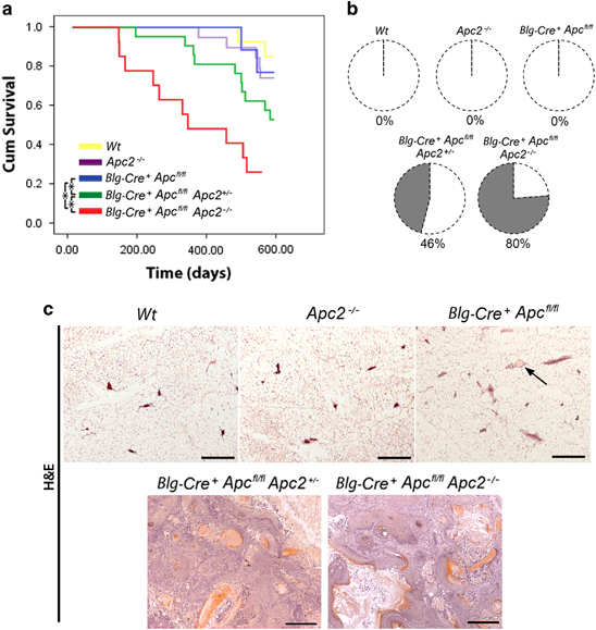
Figure 4.

Functional redundancies exist between Apc proteins in tumor suppression. (a) Wt (yellow line), Apc2−/−(purple line), Blg-Cre+Apcfl/fl (blue line), Blg-Cre+Apcfl/flApc2+/−(green line) and Blg-Cre+Apcfl/flApc2−/− (red line) mice were aged and culled upon signs of ill health (Kaplan–Meier survival curves). Apc2−/− and Blg-Cre+Apcfl/fl mice displayed no differences in survival compared with Wt mice (log-rank test, P>0.32, n⩾11). Both Blg-Cre+Apcfl/flApc2+/- and Blg-Cre+Apcfl/flApc2−/− mice displayed reduced survival compared with other genotypes (asterisks mark statistically different comparisons, P⩽0.01, log-rank test, n⩾11). (b) Mice were examined at time of death. Although Wt, Apc2−/− and Blg-Cre+Apcfl/fl mice displayed no signs of mammary pathology, 46% of Blg-Cre+Apcfl/fl Apc2+/- and 80% of Blg-Cre+Apcfl/flApc2−/− mice exhibited mammary tumors. (c) Representative images of H&E-stained mammary gland sections taken from aged mice. No lesions were found in Wt, Apc2−/− or Blg-Cre+Apcfl/fl glands, however, small infrequent intraductal aggregates of ghost cells were present in Blg-Cre+Apcfl/fl tissue (arrow). The majority of mammary tumors from Blg-Cre+Apcfl/fl Apc2+/- and Blg-Cre+Apcfl/flApc2−/− mice were classified as invasive carcinomas with squamous differentiation (scale bar, 1mm).
