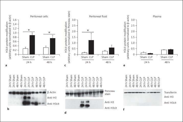
Fig. 1.
H3cit protein modification is present after CLP. a, b The H3cit protein modification is highly abundant in cells collected from the peritoneal cell lysates collected 24 h after CLP. At 48 h after CLP there is still an increase in H3cit protein modification. c, d Fluid collected from the peritoneal cavity is also increased in H3cit protein modification at 24 h after CLP, while levels decrease after 48 h. e, f At both time points, the H3cit protein modification was undetectable in the bloodstream. H3cit protein was then normalized to either β-actin for the cell lysates or to the Ponceau-stained membrane that demonstrated equal loading in the peritoneal fluid. Sham, n = 4; CLP, n = 5–6 per group. * p < 0.05, one-way ANOVA.

