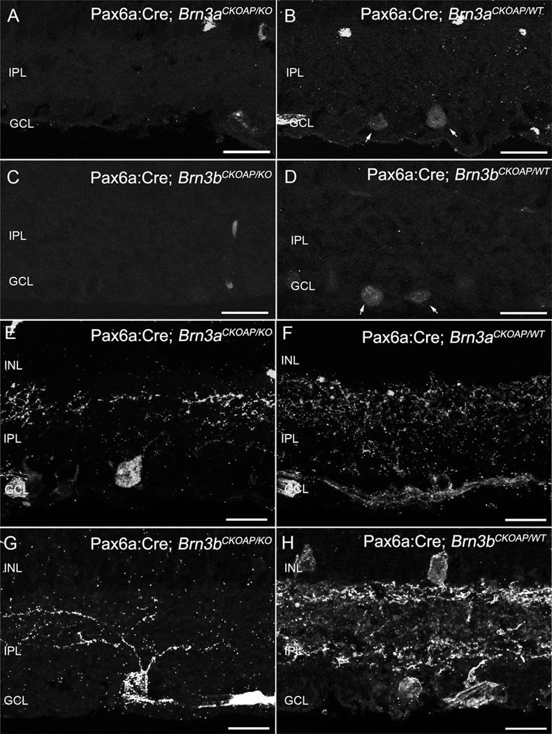
Figure 2.
Ganglion cells. Immunostaining for Brn3a and Brn3b transcription factors shows nuclei of cells located in the GCL layer (A–D). Immunoreactive nuclei are absent from the retinas of KO mice (A,C). Inner plexiform layer (IPL) of Brn3K_O mice (E,G) and of WT control (F,H) immunostained for AP revealing global Brn3a- or Brn3b–positive GCs dendritic arbors. For both KOs (D), loss of dendrites and decreased staining in the IPL are evident. A larger decrement in the number of GCs can be appreciated in the case of the Pax6α:Cre; Brn3bCKOAP/KO (G). Scale bar = 20 µm in A–H.

