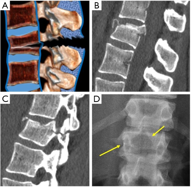
Figure 5.

Flexion-distraction fractures. (A) Sagittal scheme. (B) Sagittal CT showing interspinous widening and the horizontal fracture of the posterior arch (C). (D) Radiograph showing the empty body sign and the horizontal fracture of the pedicle (arrows).
