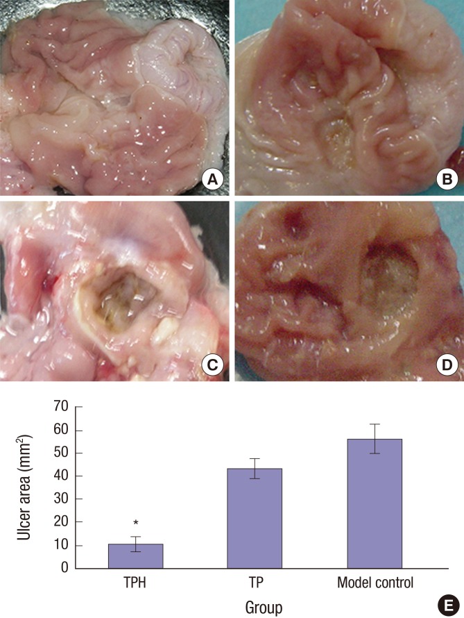
Fig. 4.
Gross observation of gastric mucosa on day 21 after induced ulcer treatment.
(A) Normal control group: the gastric mucosae from normal rats were smooth and intact with no ulceration. (B) TPH treatment group: treated with (1 × 10⁹) cfu TPH for acetic acid–induced gastric ulcer rats by gavage, the gastric mucosae of the TPH-treatment group were similar to the normal rats. The gastric mucosal lesions were smaller and shallow, and the regenerative granulation tissues were observed in ulcers. (C) Model control group: treated with 0.5-mL1.19M NaHCO3 for acetic acid–induced gastric ulcer rats by gavage. (D) TP treatment group: treated with 1 × 10⁹ cfu TP for acetic acid–induced gastric ulcer rats by gavage. In the model control and TP groups, enlarged, deepened ulcers with severe adhesions to adjacent tissues were observed, the mucosa underwent necrosis, ulcerations, and had a dark yellowish membrane-like coating. The adjacent mucosa had obvious hyperemia and edema. (E) The statistical results of the mean ulcer area size (mm2). The size of the ulcer area in 21 days after TPH treatment was smaller than that in the other two groups.
TPH = attenuated Salmonella typhimurium strain carrying a eukaryotic expression vector encodes the human HGF gene, TP = attenuated Salmonella typhimurium strain with a eukaryotic expression vector.
*P < 0.01, TPH group vs. TP and control groups.

