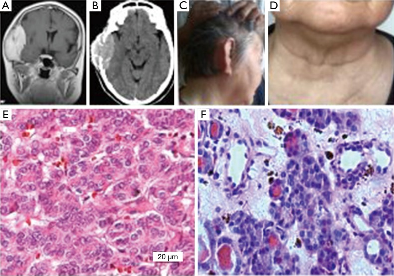
Figure 2.
Example 2: (A,B) were preoperative brain MRI and CT respectively, the showed high signal of tumor shadow located in the right temporal region, and the tumor grew inside and outside the head and destroyed the skull; (C,D) were follow-up figures which respectively showed that the right tumorectomy surgery temporal region recovered well and the anterior cervical thyroid surgery region was also normal; (E,F) were the observation of pathological slice light microscopy respectively (HE ×400), which respectively showed that the head tumor was in line with the thyroid cancer, while the thyroid tumor was in line with the thyroid cell proliferative changes.

