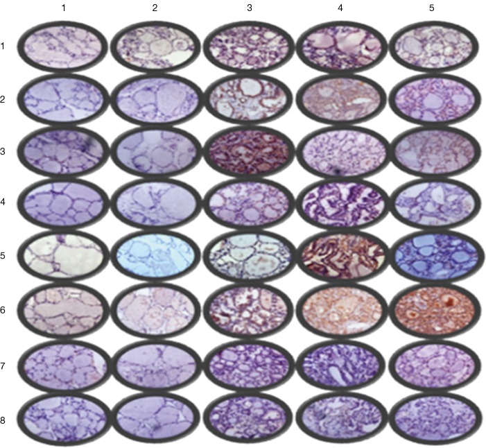
Figure 3.
Immunohistochemistry light microscopy findings (partial ×400): row 1–5 were in situ normal thyroid gland, in situ nodular goiter, in situ papillary thyroid carcinoma, metastatic thyroid cancer and ectopic thyroid cancer, respectively; column 1–8 were PI3K, AKT, mTOR, BRAF, ERK, MMP-9, IKK, and NFκB, respectively.

