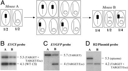
Fig. 6.
Progeny of a TARGET × DELETER-EPI cross. (A) Schematics of the parasite cross. In mouse A, the two clones are mixed in equal proportions, one having the integrated TARGET locus (black box) and the other carrying the Flp gene on an episome (open box on a circle). After transmission of the parasite mixture to mosquitoes, nuclei fusion during cross-fertilization should transmit the episome to the nuclei containing the TARGET locus. Assuming similar frequencies of self- and cross-fertilizations and stable maintenance of the episome in parasites resulting from cross-fertilizations, then 1/4 of the emerging sporozoites, as well as RBC stages in mouse B, are expected to carry both the TARGET locus and the episome-borne Flp. Episomes can be lost (+/-) during parasite multiplication in the oocyst, liver, and RBC in mouse B. Boxes indicate parasitic cells, and ellipses indicate nuclei. (B) Southern hybridization of the parasite mixture TARGET + DELETER-EPI collected from mouse A and mouse B by using a CS probe. The probe shows a similar intensity in mice A and B of the EcoRV (E5) fragments corresponding to the WT CS (4.1 kb) and the TARGET + TARGET/Exc loci (5.3 kb) (Fig. 3B). (C) Southern hybridization of the parasite mixture TARGET + DELETER-EPI collected from mouse A and mouse B by using a GFP probe. The probe shows a similar intensity in mice A and B of the EcoRV (E5) fragments corresponding to the TARGET (5.7 kb) and the TARGET/Exc (4 kb) loci (Fig. 3B). (D) Southern hybridization of the parasite mixture TARGET + DELETER-EPI collected from mouse A and mouse B by using a plasmid pUC probe. The probe detects a HincII (H2) fragment of 4.2 kb, corresponding to the TARGET or TARGET/Exc locus, in mouse A and mouse B. In contrast, the probe detects a HincII fragment of 5.3 kb (corresponding to the episome, not shown) in mouse A that is absent in RBC stages from mouse B.

