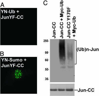
Fig. 5.
Specificity of Jun ubiquitination by Itch in living cells and cell extracts. (A and B) YN-Ub and JunY170F-CC (A) or YN-SUMO1 and JunY170F-CC (B) were expressed in COS-1 cells, and fluorescence images were acquired 36 h after transfection. (C) The proteins indicated above each lane were expressed in HEK293T cells. Cell extracts were precipitated with anti-Jun antibody, and equal amounts of the proteins were analyzed by Western blot analysis with anti-Myc antibody to detect ubiquitinated proteins (Upper). The membrane was reprobed with anti-Jun antibody to confirm that equal amounts of Jun were loaded in each lane (Lower).

